Uterine Fibroids
Uterine fibroids are common non-cancerous growths of muscle that form within the muscular wall of the uterus (womb). Fibroids occur in up to 7 out of 10 women by the time they are aged 50.
Fibroids (also known as uterine fibromyomas, leiomyomas or myomas) may grow in different layers of the uterus.
Fibroids can be located within the muscle (intramural); other types grow in the outer muscle layer (subserosal) or are inside the cavity of the uterus (submucosal).
Fibroids can vary in size, ranging from the size of a pea to the size of a rock melon or larger.
- heavy or prolonged periods
- painful periods
- anaemia or iron deficiency (due to heavy periods), and you may feel:
- tired
- dizzy
- frequent passing of urine
- a pressure sensation on the bladder, bowel or back and/or feeling of incomplete emptying of bladder or bowel
- lower back pain
- swelling in the abdomen
- painful intercourse (dyspareunia)
Bleeding in between periods is not common but can sometimes happen. In rare instances, a fibroid may become cancerous and this is called a sarcoma.
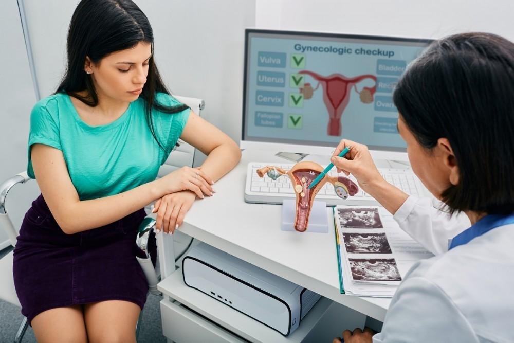
A diagnosis of fibroids may be made during a vaginal examination, ultrasound or during surgery for other conditions.
Medical Treatment Options:
- pain relief, including anti-inflammatory medications
- Tranexamic acid – a medication that reduces heaviness of bleeding by slowing blood clot breakdown in the uterine lining
- Mirena IUD – this T-shaped, contraceptive device is inserted into the uterus, and slowly releases a hormone called a progesterone, thereby reducing heavy bleeding
- Combined oral contraceptive pill – may reduce bleeding
- GnRH agonist – an artificial hormone used to prevent natural ovulation. May be used to shrink fibroid before a planned operation. Not recommended for longer term use because of side effects such as osteoporosis (bone thinning), and fibroid regrowth when the treatment is stopped.
- Iron replacement – either by oral tablets or an intravenous infusion
Surgical Treatment Options:
- Uterine artery embolisation (UAE) – small, sterile particles are injected into the uterine artery to reduce the blood supply to the fibroid, under sedation or general anaesthesia. This can make the fibroid shrink by a third or half its size.
- Hysteroscopic myomectomy – A hysteroscope is used under general anaesthesia to cut out a submucosal fibroid that is partially or completely inside the cavity of the uterus
- Myomectomy – a surgical procedure performed either laparoscopically or robotically to remove the fibroid
- Hysterectomy – removal of the uterus, either laparoscopically or robotically